
Cartilage Injury
Our Expertise
Shoulder Injuries
Recurrent Shoulder dislocation
Rotator Cuff Tear
Injuries around Shoulder
Slap Tears
Knee Injuries
ACL Injury
PCL Injury
Meniscus Injury
MPFL/MCL/LCL Injury
Cartilage Injury
Sport Injuries
ATFL Tear
Tennis Elbow
Golfer Elbow
Hip Labrel Tear
Treatment and Procedures
Shoulder Arthroscopy
Shoulder Replacement
Knee Arthroscopy
Knee Replacement
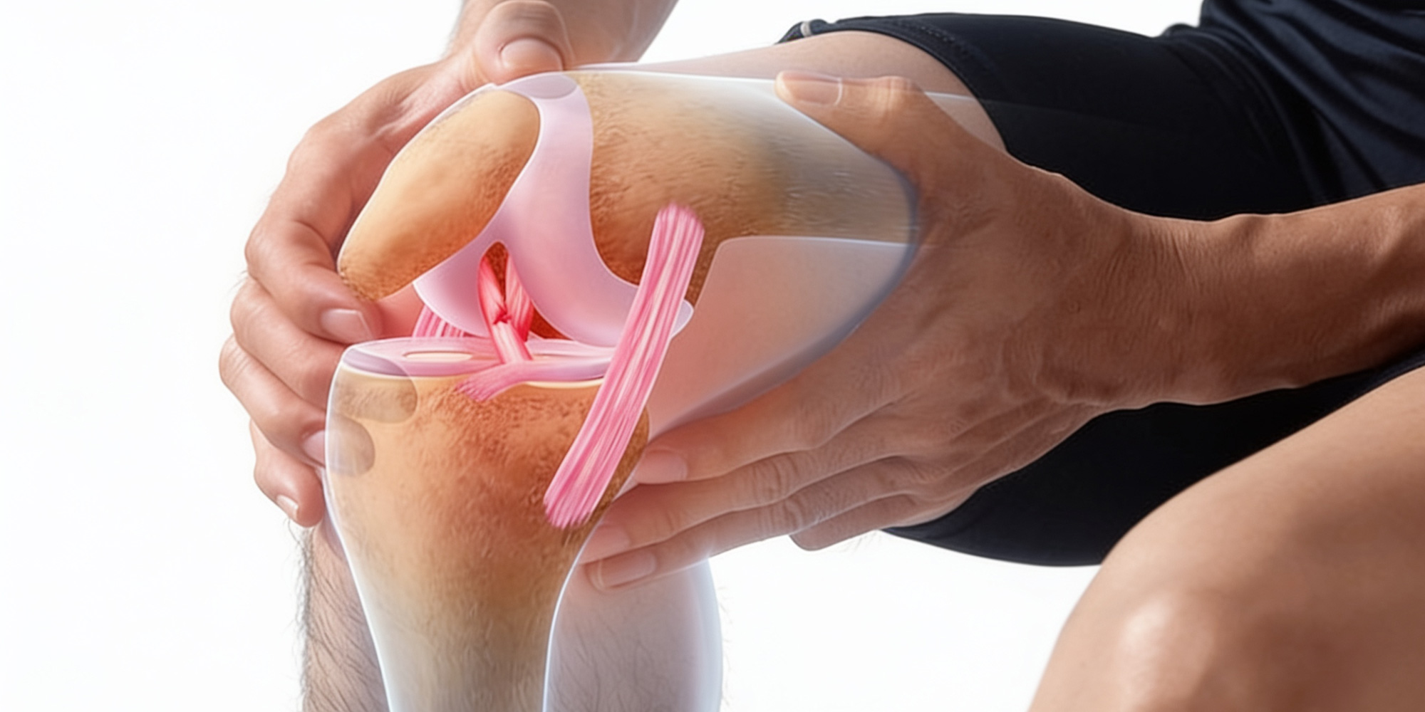
Every time you walk, run, or climb a flight of stairs, your knee joint glides smoothly because of a thin, glass-like layer called articular cartilage that coats the ends of your thigh bone, shin bone, and kneecap. This tissue has no nerve supply of its own, no significant blood flow, and a very limited capacity to heal — which is exactly what makes cartilage injuries so important to catch early and treat correctly.
A cartilage injury occurs when this smooth surface is damaged — through a sudden traumatic event, repetitive overuse, or the slow degeneration that comes with age. Unlike a bruise or a muscle strain, cartilage doesn’t repair itself the way other tissues do. Left unmanaged, even a small area of damage can progressively wear down, exposing the underlying bone and accelerating the path toward osteoarthritis.
At Jaipur Sports Doc, Dr. Abhishek Saxena focuses on identifying cartilage damage early and matching treatment precisely to the size, depth, and location of the injury — with the goal of preserving your natural joint surface for as long as possible.
What Causes Cartilage Damage?
Traumatic injury
A direct blow to the knee, an awkward twisting fall, or a sports collision can shear off or crack a section of cartilage in an instant. These injuries are common in football, basketball, and other contact or pivoting sports, and are often seen alongside ligament tears such as ACL injuries.
Repetitive Overuse
thletes and physically active individuals who place repeated high loads through the knee — runners, jumpers, and weightlifters, for example — can gradually wear down cartilage over months or years, even without a single defining injury.
Degenerative Wear
As cartilage ages, it naturally becomes thinner and less resilient. Combined with prior injuries, excess body weight, or joint misalignment, this gradual thinning is one of the leading pathways to knee osteoarthritis.
Osteochondritis dissecans
In some younger patients, a segment of cartilage and the bone beneath it can lose its blood supply and begin to separate from the surrounding joint surface, sometimes requiring specific surgical treatment of its own.
Recognizing the Symptoms
Cartilage injuries don’t always cause the sharp, obvious pain associated with a ligament tear — which is part of why they’re frequently underdiagnosed. Watch for:
- Deep, aching knee pain, often worse with activity and better with rest
- Swelling that develops after exercise or activity
- A catching, grinding, or grating sensation within the joint
- Stiffness after periods of sitting or inactivity
- A feeling of the knee locking or “giving way” if a loose fragment is present
- Pain that seems disproportionate to the apparent severity of an injury
Because cartilage has no nerve endings, symptoms often come from secondary inflammation or mechanical irritation rather than the cartilage damage itself — which means the injury can be more advanced than the pain alone would suggest.
How We Diagnose a Cartilage Injury
A thorough clinical examination assesses swelling, range of motion, joint alignment, and specific signs that point toward cartilage involvement rather than a purely ligament or meniscus problem. X-rays help rule out fractures and assess joint space and alignment, while MRI remains the most valuable tool for visualizing the cartilage layer directly — revealing the size, depth, and exact location of the defect, and whether the underlying bone is also involved.
In some cases, the full extent of a cartilage injury only becomes clear during arthroscopic examination, where Dr. Saxena can directly visualize and probe the joint surface to grade the damage accurately.
Treatment Options
Non-Surgical Management
For smaller or milder cartilage injuries, especially where symptoms are manageable, treatment often begins conservatively:
- Activity modification to reduce repetitive loading on the damaged area
- Physiotherapy to strengthen the surrounding muscles, improve joint mechanics, and reduce stress on the cartilage
- Weight management, where relevant, to reduce overall load on the knee
- Injections, including options aimed at reducing inflammation and supporting joint lubrication, in select cases
Microfracture Surgery
For focal, full-thickness cartilage defects, microfracture involves creating tiny openings in the underlying bone to stimulate a healing response, encouraging the growth of new (though structurally different) repair tissue over the damaged area. It’s a minimally invasive, arthroscopic option that works well for smaller defects.
Cartilage Restoration Procedures
For larger or more demanding defects — particularly in younger, active patients — restoration techniques aim to replace damaged cartilage with tissue that more closely resembles the joint’s original surface. These include cartilage grafting and transplantation techniques, chosen based on the size and location of the defect and the patient’s activity goals.
Addressing Underlying Contributors
Cartilage injuries rarely exist in isolation. Where malalignment, ligament instability, or meniscus damage is contributing to abnormal joint loading, Dr. Saxena addresses these factors alongside the cartilage injury itself — because treating the cartilage without correcting the underlying mechanics often leads to recurrence.
Recovery and Rehabilitation
Recovery from cartilage treatment tends to be more gradual than other knee procedures, since cartilage and its repair tissue heal slowly and require protection during the early stages. Depending on the procedure, this may involve a period of limited weight-bearing, followed by a carefully progressed physiotherapy program to rebuild strength and restore full function without overloading the healing surface. Return to sport and high-impact activity is typically staged over several months, guided by both time-based milestones and objective assessments of strength and joint readiness.
Why Choose Dr. Abhishek Saxena for Your Cartilage Injury
- Advanced diagnostic imaging and clinical assessment to accurately grade cartilage damage
- A joint-preservation philosophy that prioritizes protecting your natural cartilage wherever possible
- Minimally invasive arthroscopic techniques for both diagnosis and surgical treatment
- Treatment plans that address underlying causes — alignment, ligament stability, and meniscus health — not just the cartilage defect alone
- A structured, personalized rehabilitation program guiding you safely back to full activity
Cartilage Damage Only Gets Harder to Treat With Time
Cartilage has almost no ability to heal on its own, which means a small, manageable defect today can quietly progress into widespread joint surface damage and early arthritis if it’s left unaddressed. The earlier a cartilage injury is diagnosed, the more treatment options remain available to preserve your natural knee.